
Molecular Neuroscientist

E. Nicholas Petersen, PhD

Mechanism discovery & target validation · Method development

I am a molecular neuroscientist with five first-author publications spanning mechanosensation and circadian biology, with a track record of resolving phenotypes from their underlying molecular mechanisms. I understand that success often requires adapting or building the tool the question demands which has led me to invent four assays and instruments and build two software tools to use alongside traditional microscopy, molecular biology, and biochemistry techniques. I am an expert in super-resolution imaging, recombinant protein production and purification, and quantitative experimental design across a diverse range of topics.

E. Nicholas Petersen
Postdoctoral Associate, Rosbash Laboratory — Howard Hughes Medical Institute / Brandeis University
8
h-index
5
First-author publications
6
Assays, instruments and software built
1
Patent application drafted

Capabilities

How I work

End to end mechanism discovery

I design and use techniques at every level between a predicted structure and a measured phenotype: computational prediction, structure-guided mutagenesis, recombinant protein production, quantitative imaging, transgenic animal work, and animal behavior. This has been applied across both postdoctoral and graduate work to elucidate novel mechanisms of circadian biochemistry and mechanotransduction.

Assay and instrument development

I often built new assays and instruments when existing methods could not answer the question including a live-cell real-time enzyme-coupled PLD activity assay (patent application drafted; deployed across five papers), a temperature-controlled rapid shear-fixation apparatus (±0.1 °C, <10 s), sub-second reconstructed live-cell 3D-dSTORM, and the VAAPR anesthesia rig.

Quantitative microscopy

I have become an expert in multiple forms of advanced microscopy from 3D single-molecule localization microscopy (dSTORM/SMLM, ~6–26 nm precision) through SIM, Airyscan, TIRF, and laser-scanning confocal. I have extended many of these techniques to include sub-second live cell dSTORM recordings (~250 ms effective frame rate), super-resolution of tissue slices, and intact whole-mounted Drosophila brain. This is often paired with quantitative analysis (pair-correlation, DBSCAN, Imaris 3D) of the acquired images.

Analysis and training software

I am always looking for a more efficient way to do science which translates to better analysis as well as better techniques. To this end I developed ClockWork, co-developed in the Rosbash lab, to increase the throughput and statistical power of expensive circadian behavior experiments while putting the analysis in a user-friendly UI. Its modular design makes adding new analyses straightforward for the wider research community. A microscopy training tool, now in development, extends the same idea to a complex technique with the goal of simplifying the training and improving the data collected by the next generation of microscopists.

Selected work

Research and methods

All ten publications →
Figure from Petersen et al., bioRxiv 2025
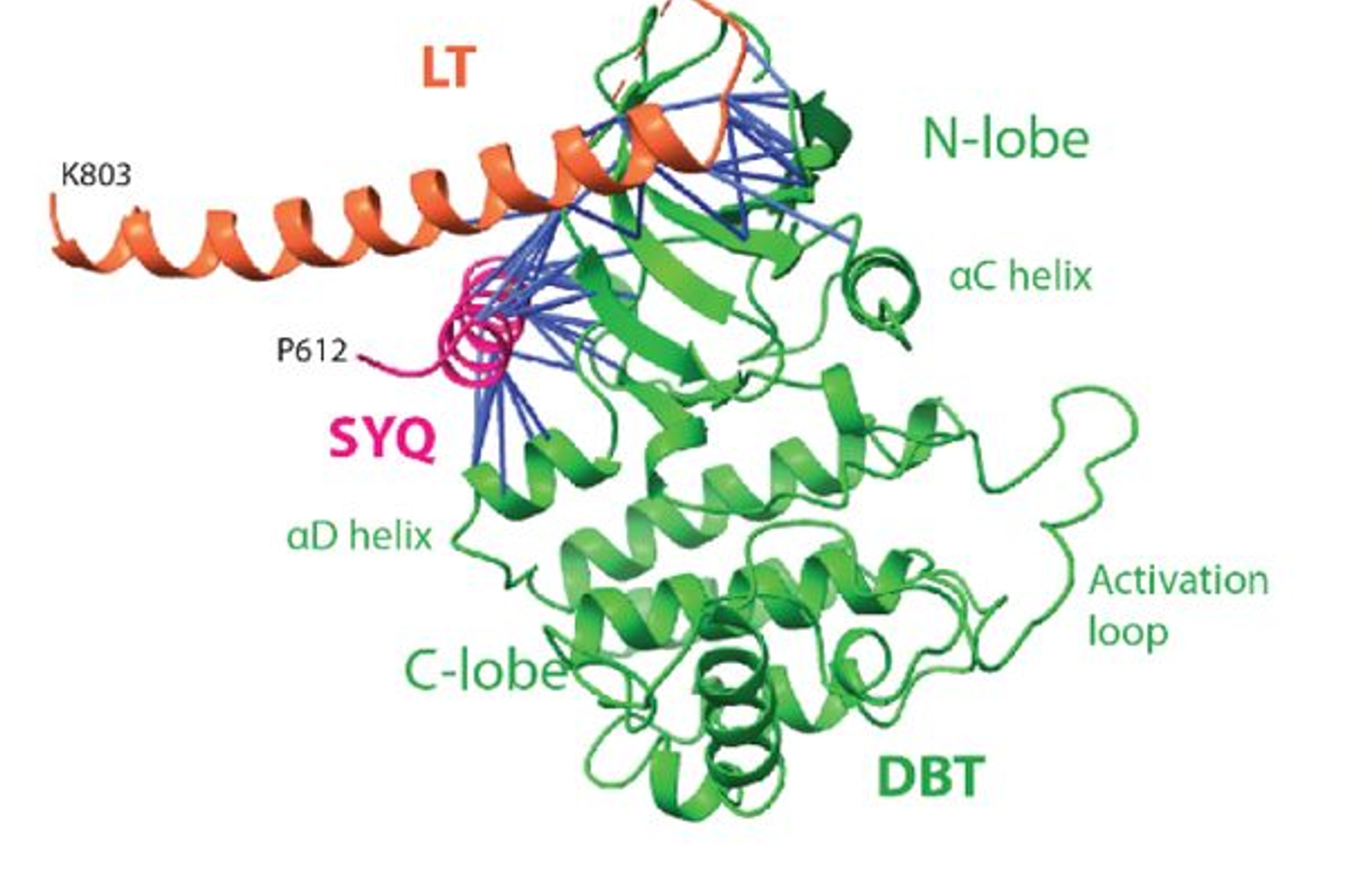
AlphaFold2-multimer model of the PER–DBT complex shows the orientation of the LT (orange) and the SYQ  (magenta) helices of the Period protein in relation to the Drosophila  kinase Doubletime.

bioRxiv preprint · 2025 · First author

The dramatic impact of the PER–DBT interaction on circadian timekeeping and temperature compensation

The circadian clock relies on phosphorylation-dependent timers to dictate period length and ensure temperature compensation. Using AlphaFold2-multimer, we predicted the interaction interface between PERIOD (PER) and DOUBLETIME (DBT/CK1). Structure-guided point mutations designed at this interface produced the longest circadian periods ever recorded in Drosophila (>48 hours) and revealed a novel, unexpected mechanism of temperature compensation driven by PER-DBT interface dynamics.
Figures, methods and PDF →

eLife · 2023 · First author

Mechanical activation of TWIK-related potassium channel by nanoscopic movement and rapid second messenger signaling

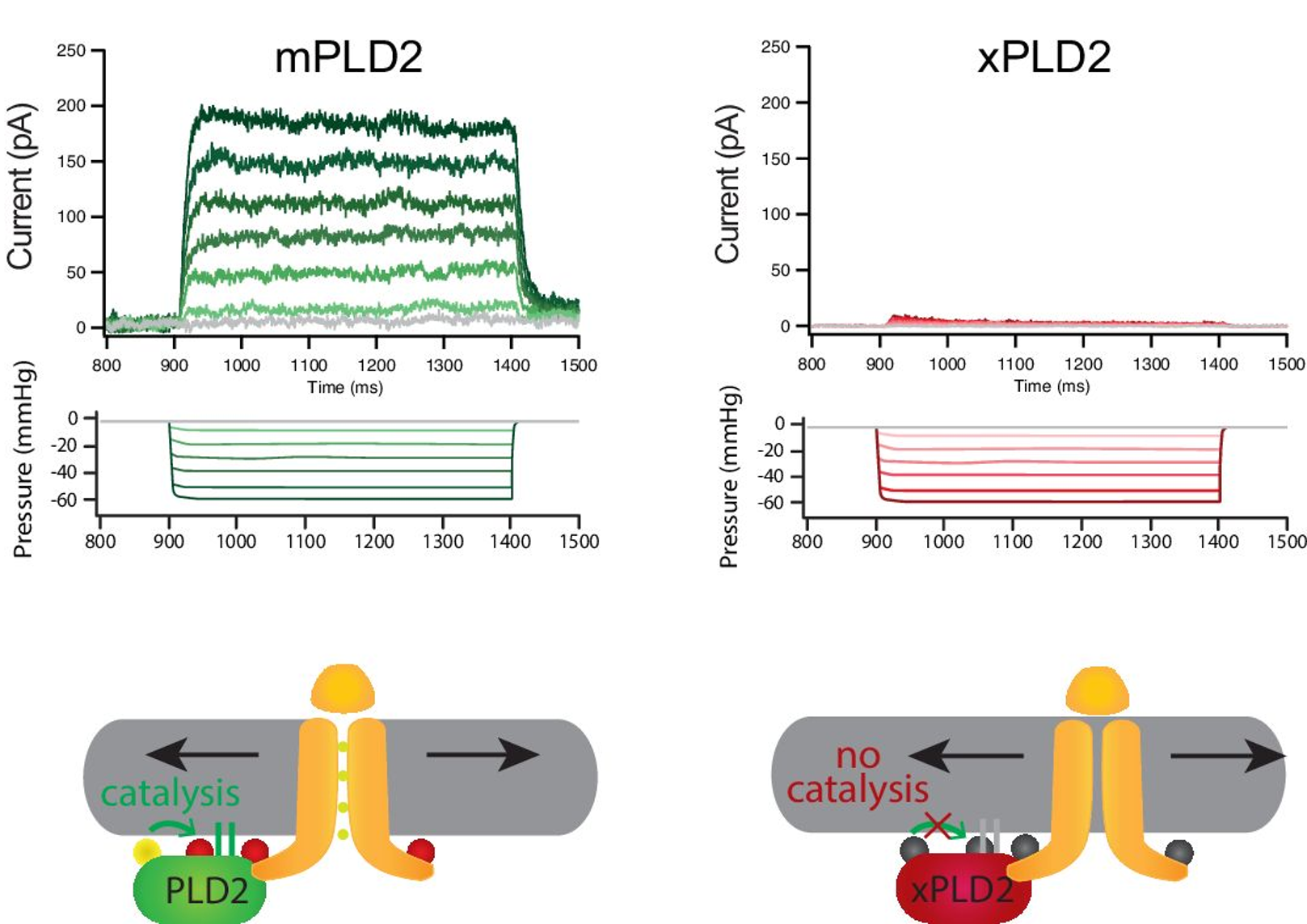
Mechanosensory ion channels like TREK-1 respond to physical force, but the molecular transducing events upstream of gating remain debated. Here we demonstrate that mechanical shear stress induces rapid, nanoscopic movement of phospholipase D (PLD2) into ordered lipid domains (GM1 rafts), producing localized phosphatidic acid (PA) that directly gates TREK-1 channels.

Figures, methods and PDF →

Figure from Petersen et al., eLife 2023
Pressure-clamp recordings of TREK-1 currents show that the mechanosensitivity of the ion channel is largely dependent on the activity of the coupled enzyme, PLD.
Live-cell enzyme-coupled PLD activity assay
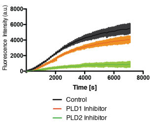
The PLD assay running in live cells. Fluorescence accumulation over time in untreated cells (black) shows the amount of PLD activity. The ability to measure PLD activity in live cells became a surrogate for testing multiple methods of membrane disruption.

Assays, instruments and software

Multiple assays, instruments, and software built to answer scientific questions

These were built when existing methods could not answer the question. A live-cell PLD activity assay deployed across five papers and drafted as a patent application, a temperature-controlled rapid shear-fixation apparatus, sub-second live-cell 3D-dSTORM, and the VAAPR anesthesia rig.

All six inventions →

Research experience

Career timeline

2020 — Present

Postdoctoral Associate — Rosbash Laboratory

Howard Hughes Medical Institute / Brandeis University, Waltham, MA

Identified the PER–DBT protein interaction as a determinant of circadian period and temperature compensation, producing the longest-period circadian mutants on record and a previously undescribed compensation mechanism. Built a structure-to-phenotype discovery workflow spanning AlphaFold2 prediction, structure-guided mutant design, recombinant protein biochemistry, transgenic animals, and quantitative behavioral/molecular readout. Currently developing this into an in silico pipeline of discovery using AlphaPulldown. Co-developed ClockWork, an open-source circadian analysis package.

2012 — 2019

Graduate Student, then Postdoctoral Fellow — Hansen Laboratory

The Scripps Research Institute, Jupiter, FL

Established disruption of ordered lipid nanodomains as a mechanism of mechanical and anesthetic activation of TREK-1 potassium channels. First author on three publications, co-author on three more. Developed a live-cell, real-time enzyme-coupled PLD activity assay used across four publications, and applied 3D single-molecule super-resolution imaging across live cells, fixed cells, mouse brain slices, and whole Drosophila brain.

2010 — 2012

Research Assistant — Buskirk Laboratory

Brigham Young University, Provo, UT

Contributed genetic selection and radiolabeled pulse-chase experiments to the identification of nascent peptide motifs that stall translation in bacteria (PNAS, 2013).

Education

Ph.D., Chemical and Biological Sciences

The Scripps Research Institute, 2019

B.S., Biochemistry

Brigham Young University, 2012

Talks & presentations

  • Invited talk, Society for Research on Biological Rhythms
  • Invited talk, Brandeis Postdoctoral Symposium, Waltham
  • Invited talk, Society for Neuroscience, San Diego
  • Invited talk, Department of Pathology, Brigham and Women's Hospital, Boston
  • Keystone Conference, Mammalian Sensory Systems, Seattle
  • GRC, Ligand Recognition and Molecular Gating, Ventura
  • Biophysical Society Annual Meeting, New Orleans

Selected awards & funding

  • NIH T32 Neurobiology Training Grant
  • Best poster, FEBS Advanced Course on Lipid–Protein Interactions, Greece
  • Scheller Graduate Fellowship, RJ Foundation
  • Best presentation, Scripps Graduate Program Retreat
  • Richard & Helen DeVos Graduate Fellowship, Scripps
  • ORCA Research Grant, Brigham Young University
  • Mentored Research Award, Brigham Young University — awarded three times